Traumatic Brain Injury Part III: Screening and Diagnosis
Traumatic Brain Injury (TBI) is a clinical diagnosis however, no single test is able to definitively confirm the diagnosis of TBI.
In Part III of our series on Traumatic Brain Injury and Litigation, we will be discussing the screening and diagnosis process. We discussed brain anatomy and function, and the specific signs and symptoms of brain injury in Part I (found here) and Part II (found here.)
The initial process for determining if a patient has a TBI is to screen for symptoms along with a physical examination. There are many screening tools available that are used by health care professionals. Some screening tools rely on observations and physical examinations by the health care provider while others rely on information provided by the patient.
Screening tools based on health care provider observation:
The Glasgow Coma Scale (GCS) is a well-known brain injury screening tool used by clinicians and is routinely performed in the emergency room or in an acute hospital setting. It may be included in the physician’s notes and is normally included in the nurse’s flow sheets. It is a simple 15-point test that measures 3 basic objective symptoms: eye-opening, motor response, and verbal response. A score of 15 is normal and anything less than that may indicate some type of abnormal neurological functioning. Both initial and worst GCS post-resuscitation scores have correlated significantly with 1-year outcomes following severe head injury.
Additional screening tools may include the Mini-Mental Status Exam (MMSE) and Luria’s motor sequencing test. MMSE evaluates cognitive functioning, analyze the quality of the patient’s language function (socially acceptable or inappropriate, impulsive or delayed), and assesses letter and number fluency. The Luria motor sequencing test can be helpful in determining if the patient has frontotemporal brain damage. An examination of cranial nerve function, hearing function, and visual/spatial function may provide clues to the area of the brain that is affected
These screening tests might be conducted by various specialties, so a review not only of the nurse’s and doctor’s notes, but also of occupational and speech therapists, physical therapists, and psychology and social work consultations can be helpful in establishing TBI.
 Screening tools for traumatic brain injury based on the self-reporting of the patient:
- HELPS brain injury tool is a simple mnemonic for Hit on Head, Emergency Room, Lose consciousness, Problems in your daily life, and Sickness.
- The Neurobehavioral Symptom Inventory is a set of 22 questions answered by the patient which is utilized by the Department of Defense to screen military personnel for TBI.
- The Brain Injury Screening Questionnaire is widely used in mental health clinics, prisons, and communities that aid the homeless and victims of domestic abuse.
There are many other tools available and review of the individual records should include any type of self-report screening tool that has been completed by the patient.
Diagnostic Testing:
Both laboratory studies and imaging studies can provide valuable information to assist the clinician in establishing a diagnosis of Traumatic Brain Injury.
Laboratory Studies:
- S100B – Several research studies have identified a correlation with TBI with increases in this protein.
- Enolase, an enzyme, is a known marker of ischemic brain damage.
- Brain-derived neurotrophic factor (BDNF), a protein, is known to be decreased in patients with severe TBI and a poor prognosis.
- The Brain Trauma Indicator, a laboratory test recently approved by the FDA (2018), measures the levels of two proteins, UCH-L1 and GFAP. Upon brain injury, these proteins are released from the brain into the blood. If found at elevated levels, brain damage with intracranial lesions, normally otherwise only visible on a CT scan, is suggested.
- Hypo or hypernatremia (too little sodium or too much sodium) may be found in brain-injured patients
- Low magnesium levels may be found in head trauma patients
A review of the medical records for a TBI patient should include identification of abnormal laboratory studies that support the TBI diagnosis.
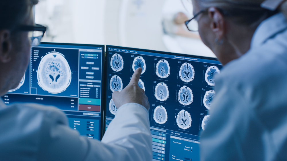
Imaging Studies:
Any accident victim or patient who has suffered a loss of consciousness should have diagnostic imaging. 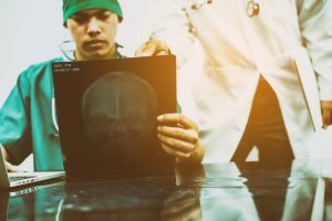
- Computerized tomography (CT) scans are the first line of imaging studies used in the emergency room for suspected head trauma. A CT scan can quickly visualize fractures and uncover evidence of bleeding in the brain (hemorrhage), blood clots (hematomas), bruised brain tissue (contusions), and brain tissue swelling. CT findings may lag behind actual intracranial damage, so that examinations performed within 3 h of trauma may underestimate injury. Repeat CT is needed when the patient deteriorates.
- Magnetic Resonance Imaging (MRI) is generally considered to be superior to CT 48 to 72 hours after the injury. MRI can detect hematomas (bruises and collections of blood) over time as the composition of blood changes. However, the majority of patients with mild brain injury show no abnormality on MRI.
- Angiography (imaging studies of blood vessels) such as computed tomography angiography (CTA) or magnetic resonance angiography (MRA) is useful to identify bleeding or hemorrhage in brain tissues.
- Diffusion Tensor Imaging (DTI) Some clinicians claim that CT and MRI do not adequately depict brain injury in TBI because they are not sensitive to detecting diffuse axonal injuries (DAI). (Axons are the long nerve fibers which connect the different brain lobes to each other). Recent advances in neuroimaging techniques, such as Diffusion Tensor Imaging, make it possible to characterize better extant brain abnormalities in mTBI. This test uses the diffusion of water molecules across brain tissues to reveal microscopic details about brain tissue architecture.
A review of a TBI case should include an analysis of a number of elements in the medical records. This includes physical examinations by doctors and nurses, screening tools administered by various specialties, laboratory studies, and imaging studies. The use of this comprehensive approach can help the attorney or paralegal to determine and/or prove the validity of a Traumatic Brain Injury claim.